


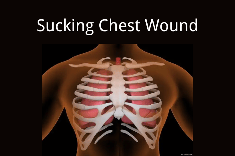
A sucking chest wound is a life-threatening medical emergency that occurs when a penetrating injury to the chest creates an open pathway for air to enter the pleural space, disrupting normal lung function. This condition, also known as an open pneumothorax, can lead to severe respiratory distress and even death if not treated promptly.
While most people may never encounter such an injury, first responders, military personnel, and outdoor enthusiasts must understand how to recognize and effectively manage it. Immediate intervention can mean the difference between life and death.
Let’s explore what a sucking chest wound is, how to identify it, and the critical steps for first aid and medical management.
Sucking Chest Wound Overview
A sucking chest wound is characterized by a penetrating injury to the chest wall that allows air to flow directly into the pleural space during inhalation. This disrupts the pressure balance required for normal lung expansion, potentially causing the affected lung to collapse (pneumothorax). The term "sucking" refers to the audible sound of air being drawn into the wound with each breath.
Causes and Mechanism of Injury
- Trauma: The most common cause is penetrating chest trauma, such as stab wounds, gunshot injuries, or impalement.
- Blunt Force Trauma: In some cases, severe blunt force can create a tear in the chest wall, leading to an open pneumothorax.
- Medical Procedures: Rarely, surgical interventions or medical procedures involving the chest can result in a sucking chest wound.
The injury creates a one-way valve effect, where air enters the pleural cavity but cannot escape, leading to increased pressure and potential tension pneumothorax.
Signs and Symptoms
- Audible Sucking Sound: A distinct sound of air entering the wound during inhalation.
- Difficulty Breathing: Shortness of breath and rapid, shallow breathing are common.
- Chest Pain: Sharp, localized pain at the site of injury.
- Visible Chest Wound: An open wound with or without blood, often accompanied by frothy blood due to air mixing with blood.
- Cyanosis: Bluish discoloration of the skin, indicating oxygen deprivation.
- Decreased Breath Sounds: Reduced or absent breath sounds on the affected side during auscultation.
Pneumothorax Types
Pneumothorax, commonly referred to as a collapsed lung, occurs when air enters the pleural space, disrupting the pressure balance necessary for lung expansion. This condition can vary in severity and presentation, depending on the underlying cause and mechanism. Understanding the different types of pneumothorax is essential for accurate diagnosis and effective treatment. This overview delves into open pneumothorax, tension pneumothorax, and the key differences between these types.
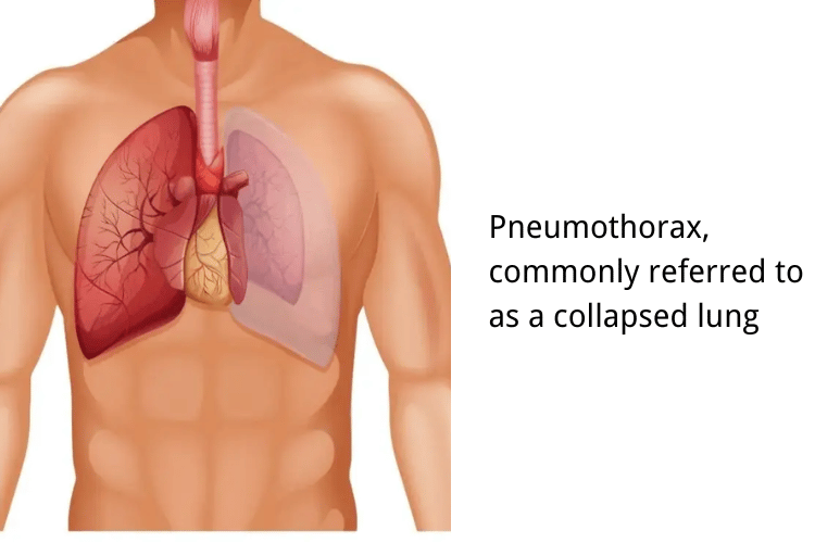
Pneumothorax, commonly referred to as a collapsed lung
Open Pneumothorax Explained
An open pneumothorax, also known as a "sucking chest wound," occurs when a penetrating injury to the chest wall creates an open channel for air to flow into the pleural cavity. This disrupts the negative pressure required for lung inflation, leading to partial or complete lung collapse. The hallmark of an open pneumothorax is the audible sound of air entering the wound during inhalation. Common causes include stab wounds, gunshot injuries, or other forms of chest trauma. Immediate intervention, such as sealing the wound with an occlusive dressing, is critical to prevent further complications.
Tension Pneumothorax: A Complication
Tension pneumothorax is a severe and life-threatening progression of pneumothorax. It occurs when air enters the pleural space but cannot escape, creating a one-way valve effect. This leads to increased intrathoracic pressure, compressing the lungs, heart, and major blood vessels. Symptoms include severe respiratory distress, hypotension, and tracheal deviation. Tension pneumothorax requires urgent decompression, typically through needle thoracostomy, to relieve pressure and restore normal function.
Differences Between Pneumothorax Types
- Mechanism: Open pneumothorax involves an external chest wall defect, while tension pneumothorax results from trapped air within the pleural cavity.
- Severity: Tension pneumothorax is more critical due to its rapid progression and impact on cardiovascular function.
- Treatment: Open pneumothorax is managed by sealing the wound, whereas tension pneumothorax requires immediate decompression to relieve pressure.
First Aid for Sucking Chest Wounds
A sucking chest wound, or open pneumothorax, is a critical medical emergency that requires immediate attention to prevent life-threatening complications such as tension pneumothorax. Proper first aid can stabilize the patient until professional medical help arrives. This guide outlines the steps for initial assessment, applying a dressing, and using a three-sided occlusive dressing to manage the injury effectively.
Initial Assessment and Response
The first step in managing a sucking chest wound is to assess the situation and ensure the safety of both the responder and the patient. Begin by checking the patient’s airway, breathing, and circulation (ABCs). Look for visible signs of a chest wound, such as an open injury with air movement or frothy blood. Listen for a sucking sound during inhalation, which is a hallmark of this condition. If the patient is conscious, reassure them and encourage shallow breathing to minimize air entry into the wound. Call emergency services immediately for advanced medical care.
Application of Dressing
To prevent further air from entering the pleural cavity, cover the wound with a sterile, non-porous material such as plastic wrap, aluminum foil, or a commercial chest seal. Ensure the material is large enough to cover the entire wound and extend beyond its edges. Apply gentle pressure to secure the dressing, but avoid pressing too hard, as this may worsen the injury. If available, use medical tape to hold the dressing in place temporarily.
Using a Three-Sided Occlusive Dressing
A three-sided occlusive dressing is a specific technique used to manage sucking chest wounds. Secure the dressing on three sides, leaving one side open to create a flutter valve effect. This allows trapped air to escape during exhalation while preventing additional air from entering during inhalation. Monitor the patient closely for signs of tension pneumothorax, such as worsening respiratory distress or tracheal deviation, and be prepared to adjust the dressing if necessary.
Management of Chest Injuries
Chest injuries, ranging from minor trauma to life-threatening conditions, require prompt and effective management to stabilize the patient and prevent complications. Proper handling during the initial stages can significantly impact the outcome. This guide focuses on key aspects of managing chest injuries, including safe patient transport, monitoring vital signs, and advanced medical interventions.
Transporting the Patient
Safe and efficient transportation is critical for patients with chest injuries. Ensure the patient is positioned to facilitate breathing, typically in a semi-upright or recovery position, unless contraindicated by spinal or other injuries. Avoid unnecessary movement to prevent exacerbating the injury. If a sucking chest wound is present, ensure the dressing is secure before transport. Emergency medical services should be contacted immediately, and the patient should be transported to the nearest trauma center for specialized care.
Monitoring Vital Signs
Continuous monitoring of vital signs is essential to assess the patient’s condition and detect any deterioration. Key parameters include respiratory rate, heart rate, blood pressure, and oxygen saturation. Look for signs of respiratory distress, such as rapid breathing, cyanosis, or use of accessory muscles. A declining level of consciousness or irregular pulse may indicate worsening conditions, such as tension pneumothorax or internal bleeding, requiring immediate intervention.
Advanced Medical Interventions
Once the patient reaches a medical facility, advanced interventions may be necessary to address the injury. These can include imaging studies like X-rays or CT scans to assess the extent of damage. Procedures such as chest tube insertion (thoracostomy) may be performed to relieve pneumothorax or hemothorax. In severe cases, surgical interventions like thoracotomy may be required to repair internal damage. Pain management and oxygen therapy are also integral to the treatment plan, ensuring patient comfort and adequate oxygenation.
Frequently Asked Questions About Sucking Chest Wound
What is a sucking chest wound?
A sucking chest wound, also known as an open pneumothorax, occurs when there is an open hole in the chest wall that allows air to enter the pleural space, disrupting the normal negative pressure inside the chest. This can lead to respiratory distress and potential lung collapse.
How does a sucking chest wound affect the lungs?
The presence of a sucking chest wound allows air to enter the chest cavity, which can result in a pneumothorax. This can lead to shortness of breath and increased pressure within the chest, ultimately causing the lung on the affected side to collapse if not managed promptly.
What are the signs of a penetrating chest injury?
Signs of a penetrating chest injury include visible holes in the chest wall, difficulty breathing, decreased breath sounds on the affected side, and signs of respiratory distress. Patients may also exhibit cyanosis or altered mental status due to lack of oxygen.
How should I cover a sucking chest wound?
To cover a sucking chest wound, apply a chest seal or a sterile dressing over the wound. Ensure that at least one side of the dressing is left open to allow air to escape from the pleural space while preventing air from entering the chest, thus avoiding tension pneumothorax.
What is the management of an open chest wound?
Management of an open chest wound involves sealing the wound to prevent air from entering the pleural cavity while allowing air to escape. Use a three-sided dressing to cover the wound, and monitor the patient for signs of respiratory distress or increased pressure within the chest.
What is the role of a chest tube in treating a sucking chest wound?
A chest tube is used to drain air, blood, or fluid from the pleural space, helping to re-establish negative pressure within the chest cavity. This treatment is crucial for patients with severe pneumothorax or hemothorax resulting from penetrating chest trauma.
What should I do if I suspect a sucking chest wound?
If you suspect a sucking chest wound, it is critical to call emergency services immediately. While waiting for help, apply a chest seal to cover the wound, monitor the patient's breathing, and position them in a way that eases their respiratory distress.
Can a sucking chest wound lead to tension pneumothorax?
Yes, a sucking chest wound can lead to tension pneumothorax if air enters the pleural space without being able to escape. This condition causes increased pressure inside the chest, which can compromise cardiovascular function and is a medical emergency requiring immediate intervention.
What imaging is used to assess a suspected sucking chest wound?
A chest x-ray is commonly used to assess for pneumothorax and other complications related to a penetrating chest injury. It can help visualize air in the chest cavity and determine the extent of lung collapse or other injuries within the chest cavity.
The Bottom Line
A sucking chest wound is a dire emergency requiring swift action. Proper sealing of the wound, monitoring for complications, and immediate transport to advanced medical care are crucial. Whether in combat, an accident, or a wilderness setting, knowing how to respond can save a life.
For first responders and medical professionals, regular training on chest trauma management is essential. For civilians, basic first aid knowledge—including recognizing a sucking chest wound—can make all the difference in a crisis.
By acting quickly and effectively, you can stabilize the patient long enough for professional help to take over, ensuring the best chance of survival.


 Login with Google
 Login with Facebook